File:Melanocytic Transformation.jpeg
Original file (600 × 647 pixels, file size: 77 KB, MIME type: image/jpeg)
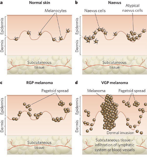
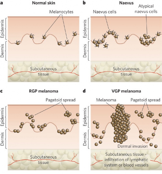
There are various stages of melanocytic lesion, each of which is marked by a new clone of cells with growth advantages over the surrounding tissues.
a, Normal skin. This shows an even distribution of dendritic melanocytes within the basal layer of the epidermis.
b, Naevus. In the early stages, benign melanocytic naevi occur with increased numbers of dendritic melanocytes. According to their localization, naevi are termed either junctional, dermal or compound. Some naevi are dysplastic, with morphologically atypical melanocytes.
c, Radial-growth-phase (RGP) melanoma. This is considered to be the primary malignant stage.
d, Vertical-growth-phase (VGP) melanoma. This is the first stage that is considered to have malignant potential and leads directly to metastatic malignant melanoma, the most deadly stage, by infiltration of the vascular and lymphatic systems. Pagetoid spread describes the upward migration or vertical stacking of melanocytes that is a histological characteristic of melanoma.
Reprinted by permission from Macmillan Publishers Ltd: Nature Publishing Group. Gray-Schopfer et al. Melanoma biology and new targeted therapy. Nature 445, 851-857(22 February 2007) [1]
File history
Click on a date/time to view the file as it appeared at that time.
| Date/Time | Thumbnail | Dimensions | User | Comment | |
|---|---|---|---|---|---|
| current | 08:04, March 19, 2015 | | 600 × 647 (77 KB) | Viraj.J.Mehta (talk | contribs) | There are various stages of melanocytic lesion, each of which is marked by a new clone of cells with growth advantages over the surrounding tissues. a, Normal skin. This shows an even distribution of dendritic melanocytes within the basal layer of the... |
You cannot overwrite this file.
File usage
The following page uses this file: