File:Medulloepithelioma MRI.jpg
From EyeWiki
Size of this preview: 799 × 274 pixels. Other resolution: 1,968 × 675 pixels.
Original file (1,968 × 675 pixels, file size: 161 KB, MIME type: image/jpeg)
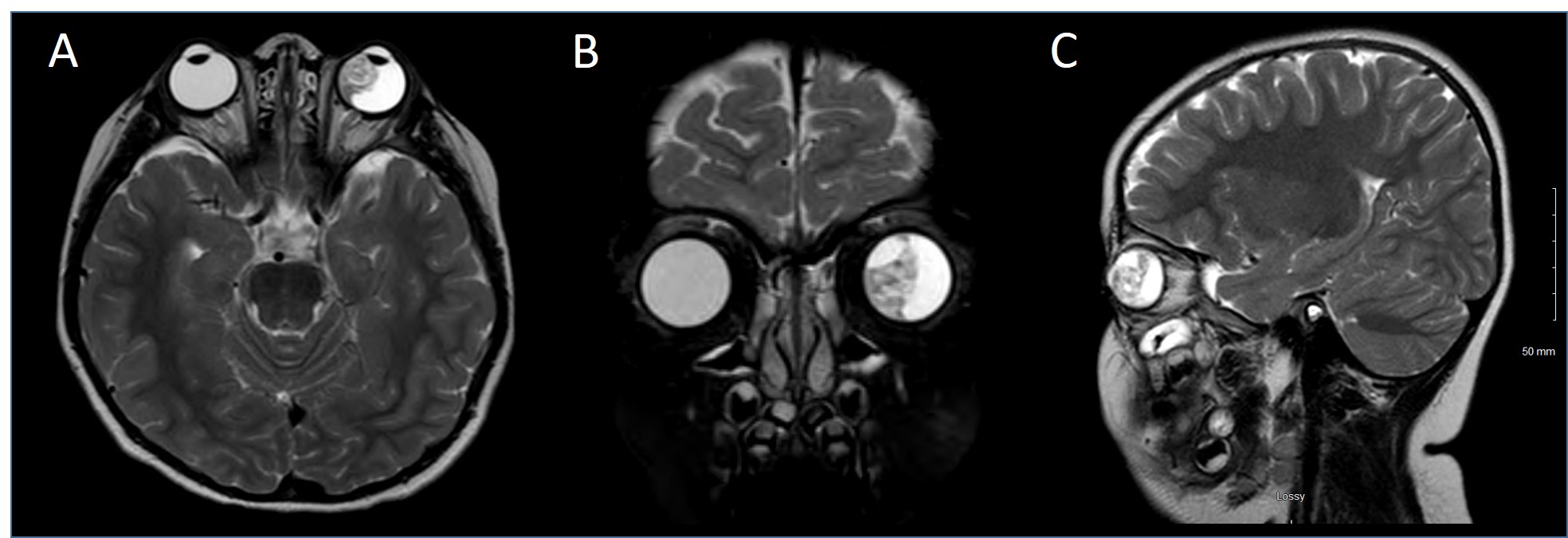
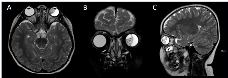
Figure 7: T2 MRI Images of a patient with A) Axial, B) Coronal and C) Sagittal sections showing an irregular soft tissue lesion within the left globe occupying nearly half of the vitreous region. The lesion is of mixed signal intensity. No obvious macroscopic retrobulbar extension is visualized.
File history
Click on a date/time to view the file as it appeared at that time.
| Date/Time | Thumbnail | Dimensions | User | Comment | |
|---|---|---|---|---|---|
| current | 17:29, November 15, 2019 | 1,968 × 675 (161 KB) | Mohammad.Sadiq (talk | contribs) | Figure 7: T2 MRI Images of a patient with A) Axial, B) Coronal and C) Sagittal sections showing an irregular soft tissue lesion within the left globe occupying nearly half of the vitreous region. The lesion is of mixed signal intensity. No obvious macr... |
You cannot overwrite this file.
File usage
The following page uses this file: