File:Medulloepithelioma - Path.jpg
Original file (1,235 × 1,093 pixels, file size: 498 KB, MIME type: image/jpeg)
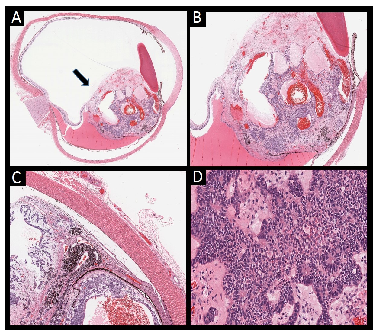
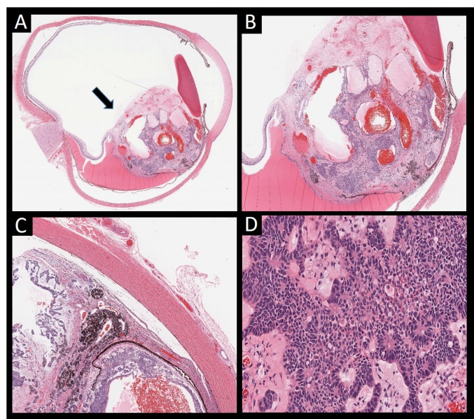
Figure 2 – Histopathological examination postenucleation of an eye with a tumor filling a large portion of the vitreous shows a: large tumor arising from the ciliary body (arrow) (A). The ciliary body mass shows basophilic cells forming ribbons and tubes with interspersed largely dilated vessels and cysts (B). Tumor cells appear to invade the choroid, with adjacent exudative retinal detachment (C). The tumor cells are round, small and deeply basophilic with some retinoblastoma differentiation. Multiple mitotic figures are present diagnostic of a malignant nonteratoid medulloepithelioma (D).
File history
Click on a date/time to view the file as it appeared at that time.
| Date/Time | Thumbnail | Dimensions | User | Comment | |
|---|---|---|---|---|---|
| current | 17:26, November 15, 2019 | | 1,235 × 1,093 (498 KB) | Mohammad.Sadiq (talk | contribs) | Figure 2 – Histopathological examination postenucleation of an eye with a tumor filling a large portion of the vitreous shows a: large tumor arising from the ciliary body (arrow) (A). The ciliary body mass shows basophilic cells forming ribbons and t... |
You cannot overwrite this file.
File usage
The following page uses this file: