File:MU Figure3.png
MU_Figure3.png (265 × 544 pixels, file size: 293 KB, MIME type: image/png)
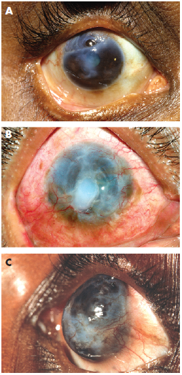
Figure 3 (A) Partial peripheral Mooren’s ulcer with a descemetocele within. Conjunctival and episcleral injection is present along with deep vessels in the base of the ulcer. A characteristic overhanging central ulcer margin is also seen. (B) Total peripheral Mooren’s ulcer with an edematous, opacified central cornea. (C) Complete Mooren’s ulcer where a fibrovascular membrane has replaced the corneal stroma. (Figure reproduced from Srinivasan, Zegans, Zelefsky, Kundu, Lietman, Whitcher, and Cunningham [3] with permission from the British Journal of Ophthalmology.)
File history
Click on a date/time to view the file as it appeared at that time.
| Date/Time | Thumbnail | Dimensions | User | Comment | |
|---|---|---|---|---|---|
| current | 20:18, August 4, 2015 | | 265 × 544 (293 KB) | Frank.S.Hwang (talk | contribs) | Figure 3 (A) Partial peripheral Mooren’s ulcer with a descemetocele within. Conjunctival and episcleral injection is present along with deep vessels in the base of the ulcer. A characteristic overhanging central ulcer margin is also seen. (B)... |
You cannot overwrite this file.
File usage
The following page uses this file: