File:Histology Apocrine Hidrocystoma.png
From EyeWiki
Size of this preview: 739 × 600 pixels. Other resolution: 1,412 × 1,146 pixels.
Original file (1,412 × 1,146 pixels, file size: 2.52 MB, MIME type: image/png)
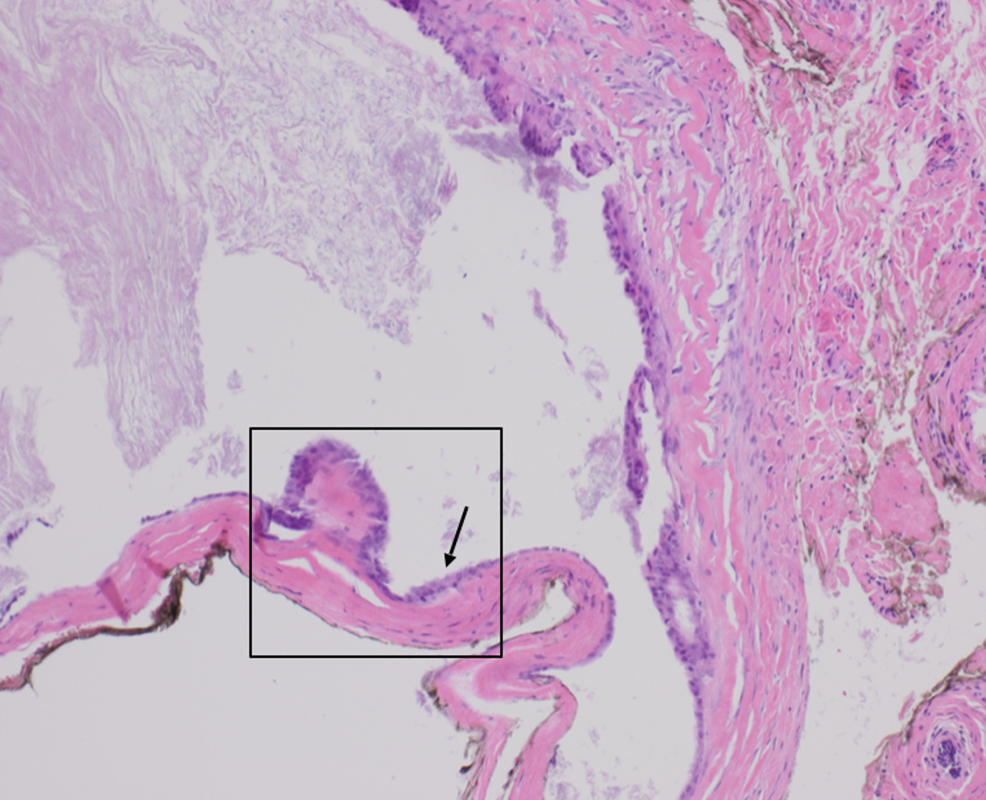
Unilocular cyst is lined by a thin layer of squamous epithelium consisting of two cell layers. (hematoxylin and eosin, original magnification x10). Pathology interpretation by Obianuju Mercy Anelo MD, Nina Krassilnik MD at UTHSC-Memphis.
File history
Click on a date/time to view the file as it appeared at that time.
| Date/Time | Thumbnail | Dimensions | User | Comment | |
|---|---|---|---|---|---|
| current | 12:18, January 8, 2022 | | 1,412 × 1,146 (2.52 MB) | Fabliha.Anbar (talk | contribs) |
You cannot overwrite this file.
File usage
The following page uses this file: