File:Hisopathology biettis.png
From EyeWiki

Size of this preview: 616 × 600 pixels. Other resolution: 717 × 698 pixels.
Original file (717 × 698 pixels, file size: 430 KB, MIME type: image/png)
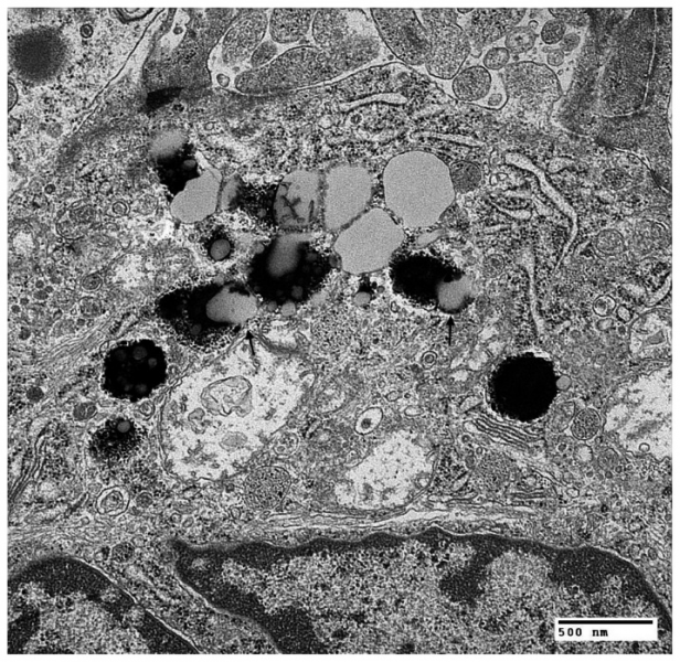
Ciliary Body Viewed With Transmission Electron Micrograph Arrow points to melanosome lipid-complex inclusions found in a pigmented ciliary epithelium (Furusato 2010).
File history
Click on a date/time to view the file as it appeared at that time.
| Date/Time | Thumbnail | Dimensions | User | Comment | |
|---|---|---|---|---|---|
| current | 15:53, July 22, 2019 | | 717 × 698 (430 KB) | Scdryden (talk | contribs) | Ciliary Body Viewed With Transmission Electron Micrograph Arrow points to melanosome lipid-complex inclusions found in a pigmented ciliary epithelium (Furusato 2010). |
You cannot overwrite this file.
File usage
The following page uses this file: