File:Figure 9 - Clinical example of a patient with diabetic macular ischemia.jpg
From EyeWiki
Size of this preview: 408 × 599 pixels. Other resolution: 595 × 873 pixels.
Original file (595 × 873 pixels, file size: 138 KB, MIME type: image/jpeg)
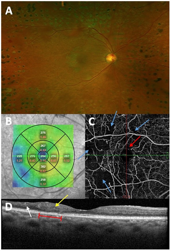
A – Fundus photo of “featureless retina.” B – En-face OCT showing central subfoveal thickness decreased to 204, suggestive of neurosensory atrophy. C – En-face OCTA confirming DMI. Red arrow – Mildly irregular FAZ outline. Blue arrows – Non-contiguous areas of macular capillary non-perfusion. D – OCT B-scan along fovea, showing signs of non-perfusion. Yellow arrow – Outer retinal disruption. White arrow – Retinal thinning. Red bracket – IS/OS junction disruption and photoreceptor damage.
File history
Click on a date/time to view the file as it appeared at that time.
| Date/Time | Thumbnail | Dimensions | User | Comment | |
|---|---|---|---|---|---|
| current | 12:53, October 20, 2021 | | 595 × 873 (138 KB) | Cris.Jacoba (talk | contribs) |
You cannot overwrite this file.
File usage
The following page uses this file: