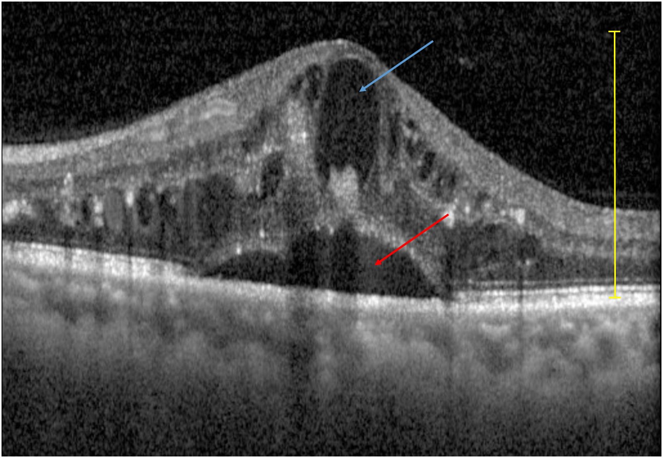
File:Figure 4 Basic Structural Changes seen in OCT with DME.jpg
From EyeWiki
Size of this preview: 800 × 553 pixels. Other resolution: 1,294 × 895 pixels.
Original file (1,294 × 895 pixels, file size: 214 KB, MIME type: image/jpeg)
Yellow bracket– Generalized retinal swelling. Blue arrow– Cystoid macular edema. Red arrow– Subretinal fluid.
File history
Click on a date/time to view the file as it appeared at that time.
| Date/Time | Thumbnail | Dimensions | User | Comment | |
|---|---|---|---|---|---|
| current | 11:55, February 23, 2022 | | 1,294 × 895 (214 KB) | Cris.Jacoba (talk | contribs) |
You cannot overwrite this file.
File usage
The following page uses this file: