File:Figure 3 ADNIV.png
From EyeWiki
No higher resolution available.
Figure_3_ADNIV.png (251 × 571 pixels, file size: 224 KB, MIME type: image/png)
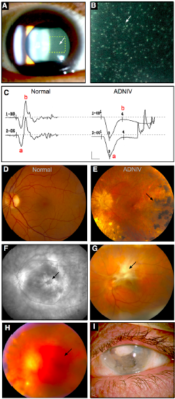
Clinical features of ANIV. A-B. demonstrating leukocytes of the vitreous. C. ERG demonstrating loss of B wave. D-E. demonstrating pigmentary degeneration of Stage 2. F. Cystoid macular edema on fluorescein angiography. G. Retinal fibrosis seen in Stage 3. H. Vitreous hemorrhage from neovascularization seen in Stage 4. I. Phthsis bulbi seen in Stage 5. (Adapted with permission from Mahajan et al 2012 PLoS Genetics)
File history
Click on a date/time to view the file as it appeared at that time.
| Date/Time | Thumbnail | Dimensions | User | Comment | |
|---|---|---|---|---|---|
| current | 12:40, February 10, 2018 | | 251 × 571 (224 KB) | Ryan.A.Shields (talk | contribs) | Clinical features of ANIV. A-B. demonstrating leukocytes of the vitreous. C. ERG demonstrating loss of B wave. D-E. demonstrating pigmentary degeneration of Stage 2. F. Cystoid macular edema on fluorescein angiography. G. Retinal fibrosis seen in Stage... |
You cannot overwrite this file.
File usage
The following page uses this file: