File:Figure 3-LASIK.jpg
From EyeWiki
Size of this preview: 800 × 187 pixels. Other resolution: 1,800 × 420 pixels.
Original file (1,800 × 420 pixels, file size: 155 KB, MIME type: image/jpeg)
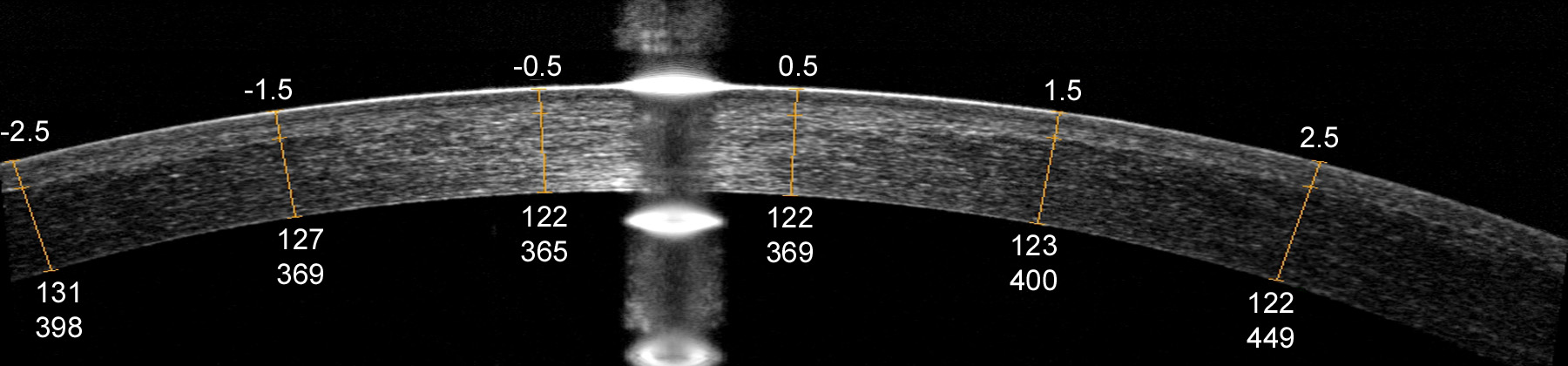
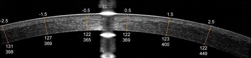
Figure 3. Optical coherence tomography (OCT) image of a LASIK flap showing measurement of the flap and stromal bed thickness. The upper numbers represent the transverse distance from the corneal vertex in millimetres. The lower numbers represent flap thickness and residual stromal bed thickness in microns, respectively.
File history
Click on a date/time to view the file as it appeared at that time.
| Date/Time | Thumbnail | Dimensions | User | Comment | |
|---|---|---|---|---|---|
| current | 12:00, June 22, 2010 | 1,800 × 420 (155 KB) | Davidhuang (talk | contribs) | Figure 3. Optical coherence tomography (OCT) image of a LASIK flap showing measurement of the flap and stromal bed thickness. The upper numbers represent the transverse distance from the corneal vertex in millimetres. The lower numbers represent flap th |
You cannot overwrite this file.
File usage
The following page uses this file: