File:Figure 2 SWM.jpg
From EyeWiki

No higher resolution available.
Figure_2_SWM.jpg (624 × 450 pixels, file size: 92 KB, MIME type: image/jpeg)
FIGURE 2: Localized Psammomatous Sphenoid Wing Meningioma stained with H&E (20x). Image demonstrates diffuse psammoma- calcifications often found at the center of the meningothelial whorls.
File history
Click on a date/time to view the file as it appeared at that time.
| Date/Time | Thumbnail | Dimensions | User | Comment | |
|---|---|---|---|---|---|
| current | 16:07, November 2, 2013 | | 624 × 450 (92 KB) | Kenneth.M.Downes (talk | contribs) | Reverted to version as of 00:05, 3 November 2013 |
| 16:06, November 2, 2013 | 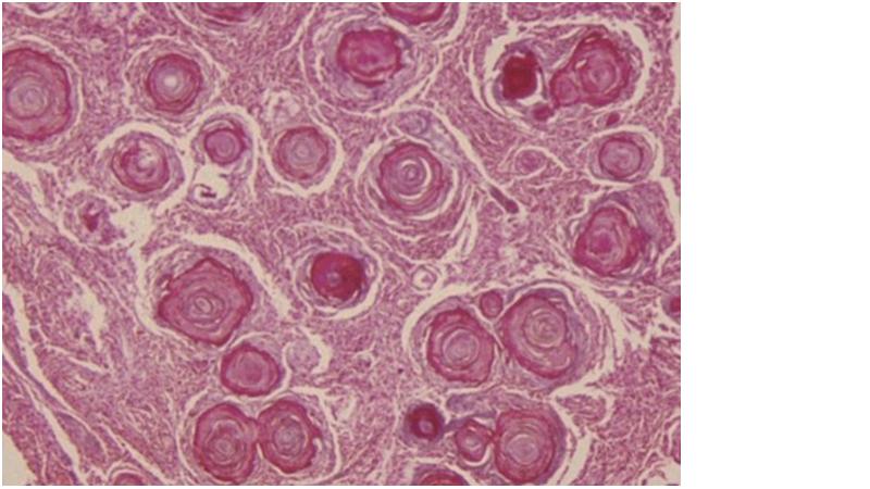 | 800 × 450 (58 KB) | Kenneth.M.Downes (talk | contribs) | Reverted to version as of 23:31, 2 November 2013 | |
| 16:05, November 2, 2013 | 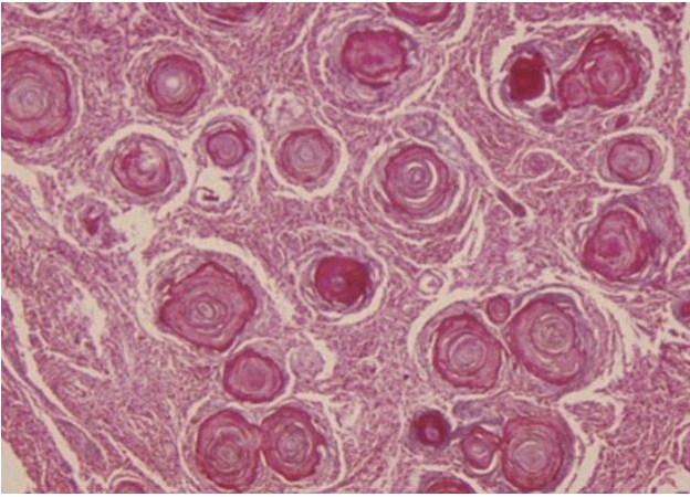 | 624 × 450 (92 KB) | Kenneth.M.Downes (talk | contribs) | ||
| 16:03, November 2, 2013 |  | 624 × 450 (92 KB) | Kenneth.M.Downes (talk | contribs) | ||
| 15:31, November 2, 2013 |  | 800 × 450 (58 KB) | Kenneth.M.Downes (talk | contribs) | FIGURE 2: Localized Psammomatous Sphenoid Wing Meningioma stained with H&E (20x). Image demonstrates diffuse psammoma- calcifications often found at the center of the meningothelial whorls. |
You cannot overwrite this file.
File usage
The following page uses this file: