File:Fig. 2B - iris diffuse mm path.jpg
From EyeWiki
No higher resolution available.
Fig._2B_-_iris_diffuse_mm_path.jpg (400 × 268 pixels, file size: 148 KB, MIME type: image/jpeg)
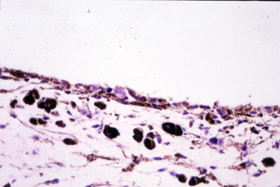
Figure 2B: Histopathologic examination reveals a heavily pigmented tumor composed of spindle B and epitheloid cells as well as melanomacrophages.
File history
Click on a date/time to view the file as it appeared at that time.
| Date/Time | Thumbnail | Dimensions | User | Comment | |
|---|---|---|---|---|---|
| current | 15:27, March 2, 2011 | | 400 × 268 (148 KB) | Martina.C.Herwig (talk | contribs) | Figure 2B: Histopathologic examination reveals a heavily pigmented tumor composed of spindle B and epitheloid cells as well as melanomacrophages. |
You cannot overwrite this file.
File usage
The following page uses this file: