File:Dome shaped macula.png
From EyeWiki
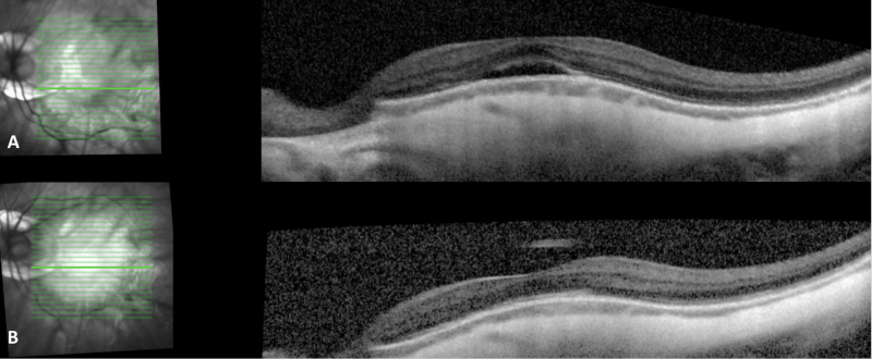
Size of this preview: 800 × 330 pixels. Other resolution: 1,908 × 788 pixels.
Original file (1,908 × 788 pixels, file size: 1.24 MB, MIME type: image/png)
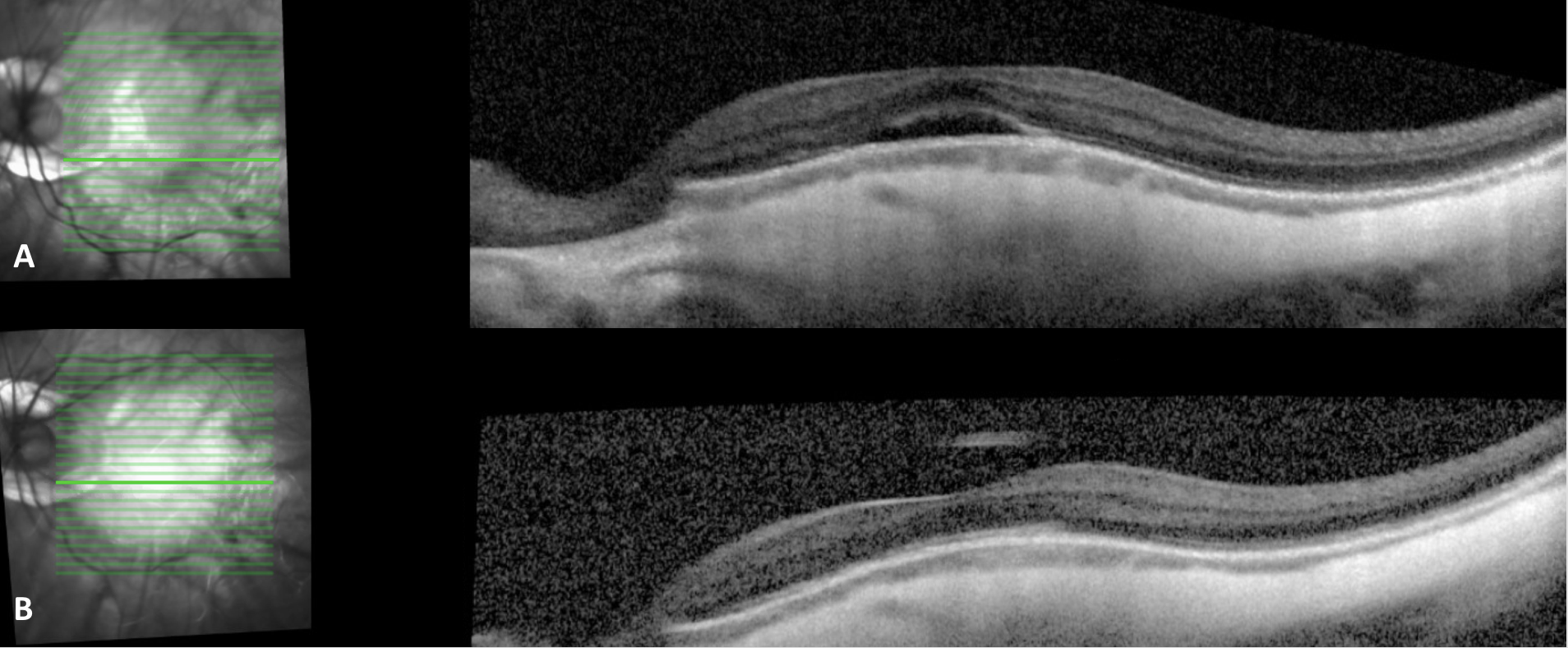
Optical coherence tomography through the macula, left eye, of a 26 year myopic (-20.00) male with A) a dome shaped appearance to the subfoveal macula with subfoveal subretinal fluid, a thin choroid, and thickened sclera. B) 3 months later, OCT demonstrates resolution of the subfoveal subretinal fluid and reconstitution of the foveal contour. Significant peripapillary atrophy can also be noted in the infrared images.
File history
Click on a date/time to view the file as it appeared at that time.
| Date/Time | Thumbnail | Dimensions | User | Comment | |
|---|---|---|---|---|---|
| current | 19:52, May 9, 2019 | | 1,908 × 788 (1.24 MB) | Danny.A.Mammo (talk | contribs) | Optical coherence tomography through the macula, left eye, of a 26 year myopic (-20.00) male with A) a dome shaped appearance to the subfoveal macula with subfoveal subretinal fluid, a thin choroid, and thickened sclera. B) 3 months later, OCT demonstr... |
You cannot overwrite this file.
File usage
The following page uses this file: