File:AS-OCT Acute Corneal Hydrops.png
Original file (1,138 × 812 pixels, file size: 835 KB, MIME type: image/png)
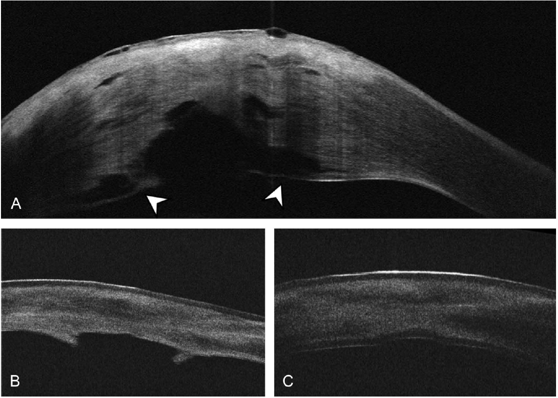
A, AS-OCT of the cornea at 10 days after presentation showing reattachment of DM. Shadowing from the stromal obscures part of the posterior cornea edema. In the visualized portion of the posterior cornea, a water cleft separates a thin layer of posterior stroma and DM, from the remaining stroma (arrowheads). B, AS-OCT 5 weeks after presentation demonstrates improving corneal edema with persistent haze, an irregular contour of the central posterior cornea, and protrusions of tissue. The contour of the central posterior cornea is irregular. C, AS-OCT 1week after ultrathin DSAEK confirms a well-attached graft.
File history
Click on a date/time to view the file as it appeared at that time.
| Date/Time | Thumbnail | Dimensions | User | Comment | |
|---|---|---|---|---|---|
| current | 13:19, May 1, 2021 | | 1,138 × 812 (835 KB) | Farida.Hakim (talk | contribs) |
You cannot overwrite this file.
File usage
The following page uses this file: