File:AIM clinical progression.jpeg
Original file (2,086 × 1,468 pixels, file size: 348 KB, MIME type: image/jpeg)
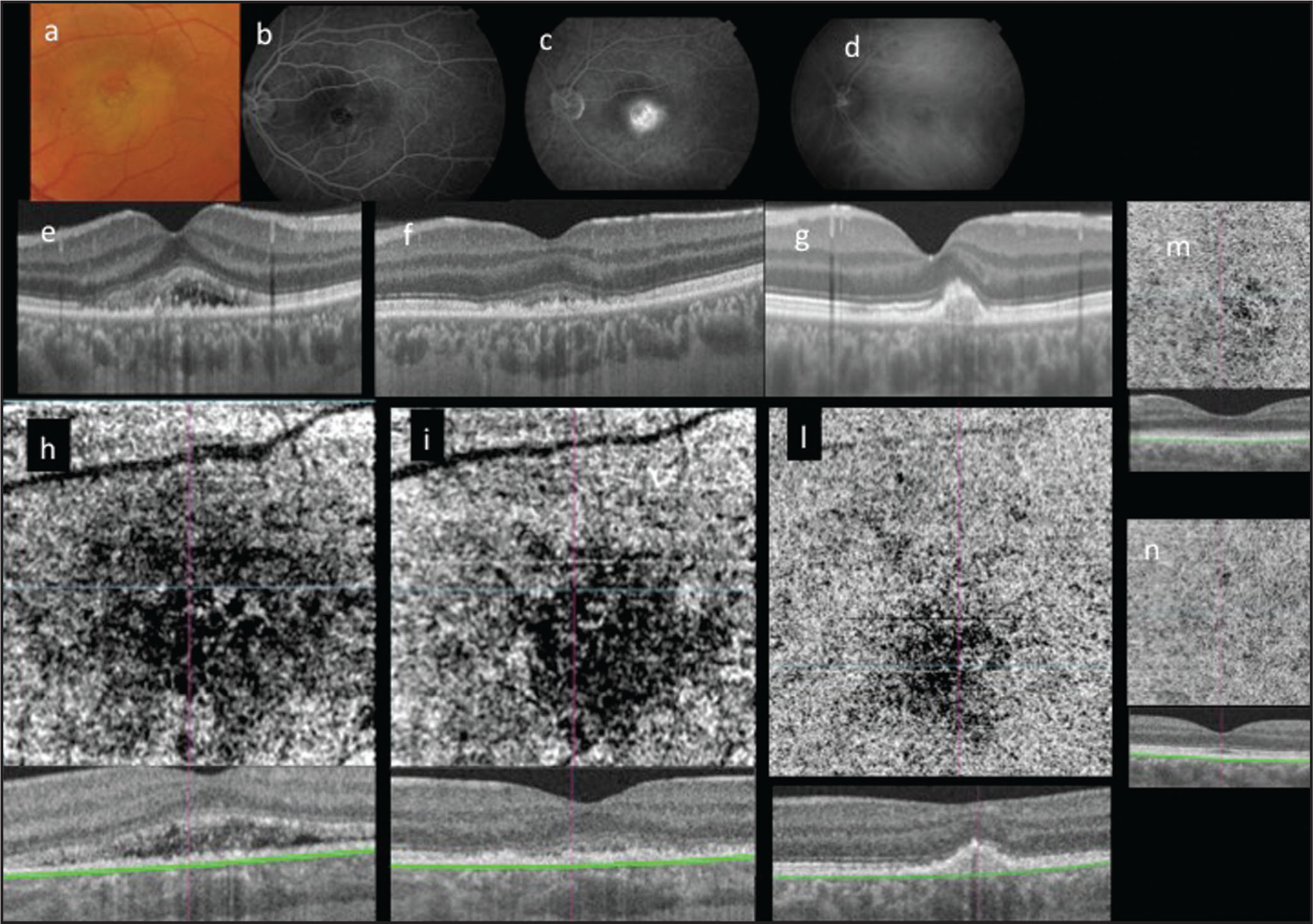
Photographs of the left eye of a patient with unilateral acute idiopathic maculopathy (UAIM) at the initial visit (A–E, H), after 4 days (F, I) and at 1 month (G, L). (A) Fundus photograph showing a yellowish lesion with an orange/dark core in the left macula. (B, C) Fluorescein angiography showing central hypofluorescence with surrounding faint, ring-shaped hyperfluorescence in the initial phase (B) and marked leakages and pooling of the dye within the subretinal space in the late phase (C). (D) Indocyanine green angiography showing a hypofluorescent lesion with some hyperfluorescence in the mid-late phase. (E) Swept-source optical coherence tomography (SS-OCT) at the initial visit (E) showing neurosensory detachment of the retina with thickening of the outer retina due to disruption and irregularity of the photoreceptor outer segment. At 4 days (F), imaging showed partial resolution of the neurosensory detachment with a better delineation of the outer retina layers. (C) At 1 month (G), showed a complete resolution of the neurosensory detachment with gradual improvement of the outer retinal layers and a juxtafoveal focal retinal pigment epithelial detachment containing hyporeflective material. SS-OCT angiography photographs taken at the level of the choriocapillaris segmentation line in the study (H, I, L), fellow (M), and control healthy (N) eyes. (H, I) Photographs showing the dark pattern and the progressive reduction compared to the normal “white noise” effect of the fellow (M) and control healthy (N) eye.
File history
Click on a date/time to view the file as it appeared at that time.
| Date/Time | Thumbnail | Dimensions | User | Comment | |
|---|---|---|---|---|---|
| current | 14:30, November 29, 2016 | | 2,086 × 1,468 (348 KB) | Jennifer.R.Gallagher (talk | contribs) | Photographs of the left eye of a patient with unilateral acute idiopathic maculopathy (UAIM) at the initial visit (A–E, H), after 4 days (F, I) and at 1 month (G, L). (A) Fundus photograph showing a yellowish lesion with an orange/dark core in the le... |
You cannot overwrite this file.
File usage
The following page uses this file: