File:AA0 Sagging Eye rads.jpeg
From EyeWiki

Size of this preview: 595 × 599 pixels. Other resolution: 2,100 × 2,115 pixels.
Original file (2,100 × 2,115 pixels, file size: 1.14 MB, MIME type: image/jpeg)
Summary
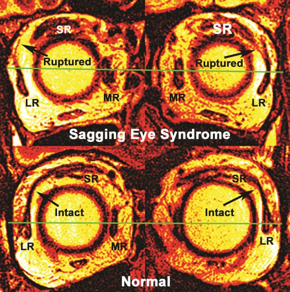
Courtesy of Joseph L. Demer, MD, PhD. Sagging eye syndrome. "This coronal magnetic resonance imaging scan demonstrates inferior “sagging” of the lateral rectus (LR) with rupture of the LR–superior rectus (SR) band bilaterally. The horizontal line depicts the center of the medial rectus (MR) muscle, which intersects the upper pole of the LR muscle"
File history
Click on a date/time to view the file as it appeared at that time.
| Date/Time | Thumbnail | Dimensions | User | Comment | |
|---|---|---|---|---|---|
| current | 16:28, January 22, 2022 | | 2,100 × 2,115 (1.14 MB) | Claudia.Prospero.Ponce (talk | contribs) | Courtesy of Joseph L. Demer, MD, PhD. Sagging eye syndrome. "This coronal magnetic resonance imaging scan demonstrates inferior “sagging” of the lateral rectus (LR) with rupture of the LR–superior rectus (SR) band bilaterally. The horizontal line depicts the center of the medial rectus (MR) muscle, which intersects the upper pole of the LR muscle" |
You cannot overwrite this file.
File usage
The following page uses this file: